Pigmented Spots: Why Laser Choice Depends on Pigment Type
Lorema Estetic
“Can you remove these sun spots?” sounds like a straightforward request. In practice, the answer depends on what the spots are, where the pigment is located and how the client’s skin responds to inflammation.
A brown mark may be a solar lentigo, post-inflammatory hyperpigmentation, melasma or a deeper pigmentary condition. These concerns can look similar but require different strategies. A laser that is suitable for a well-defined epidermal lesion may be ineffective or unnecessarily aggressive for another type of pigmentation.
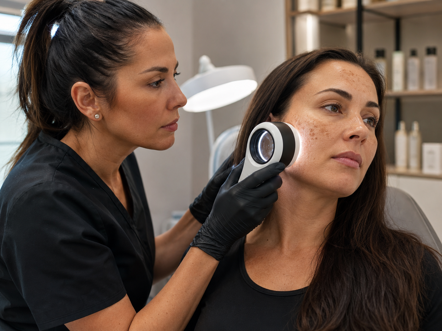
Professional treatment planning begins with three questions:
-
What caused the pigmentation?
-
How deeply is the pigment located?
-
How likely is the skin to develop an inflammatory pigment response?
Common types of pigmented spots
Solar lentigines and freckles
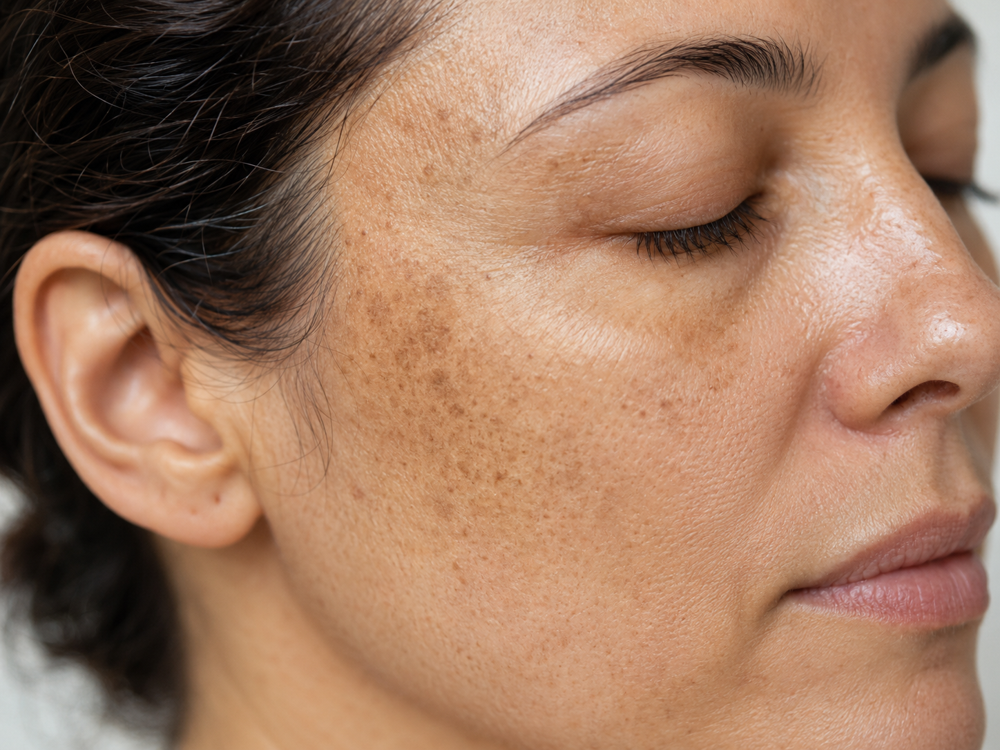
Solar lentigines often develop on areas with repeated sun exposure, including the face, hands and décolletage. Many have a strong epidermal component and may be considered for selective treatment once their benign nature is confirmed.
Removing an existing spot does not prevent further photodamage. Consistent sun protection remains essential.
Post-inflammatory hyperpigmentation
PIH can follow acne, burns, peels, trauma or other inflammatory processes. It tends to be more visible and persistent in melanin-rich skin.
The priority is to avoid creating further inflammation. Systematic reviews describe both improvement and worsening after laser-based interventions, which is why conservative settings and careful patient selection matter. See the systematic review of post-inflammatory hyperpigmentation treatments.
Melasma
Melasma commonly appears as symmetrical brown or grey-brown facial patches. It is a multifactorial condition involving light exposure, hormonal influences, genetic susceptibility and changes within the dermis and vasculature.
Aggressive energy delivery may trigger irritation, rebound pigmentation or recurrence. Laser treatment is therefore considered within a broader management plan rather than as a universal first-line solution. See the systematic review of laser therapy for melasma.
Dermal and mixed pigmentation
Grey or blue tones may suggest a deeper pigment component, although colour alone is not a diagnosis. Mixed pigmentation may involve both the epidermis and dermis and can require a staged plan or medical referral.
Why laser selection depends on the target
Pigment-selective laser treatment is based on selective photothermolysis. A chosen wavelength is absorbed by melanin, while pulse duration and energy are adjusted to limit unwanted effects on surrounding structures. The principle was introduced in the landmark paper on selective photothermolysis.
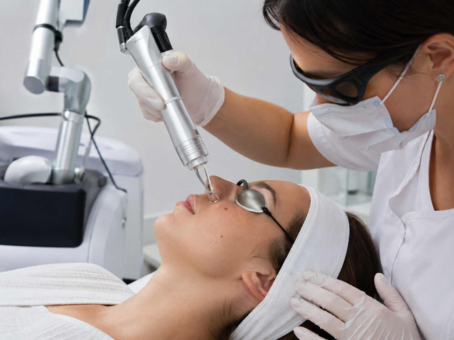
Treatment response also depends on:
-
pulse duration;
-
fluence;
-
spot size;
-
number of passes;
-
pigment depth;
-
skin phototype;
-
recent tanning;
-
previous inflammatory responses.
The useful question is therefore not “Which pigmentation laser is best?” but “Which wavelength and parameters suit this target and this skin?”
The different roles of 532 and 1064 nm
532 nm for selected superficial targets
The 532 nm wavelength is well absorbed by melanin and is used in selected protocols for benign epidermal pigment.
Epidermal melanin outside the lesion can also absorb energy, so darker phototypes, recent tanning and a history of PIH require a particularly cautious approach.
1064 nm for deeper penetration
The 1064 nm wavelength penetrates more deeply and interacts less strongly with epidermal melanin. Within Q-switched Nd:YAG systems, it can be used for selected deeper targets and carefully adapted protocols.
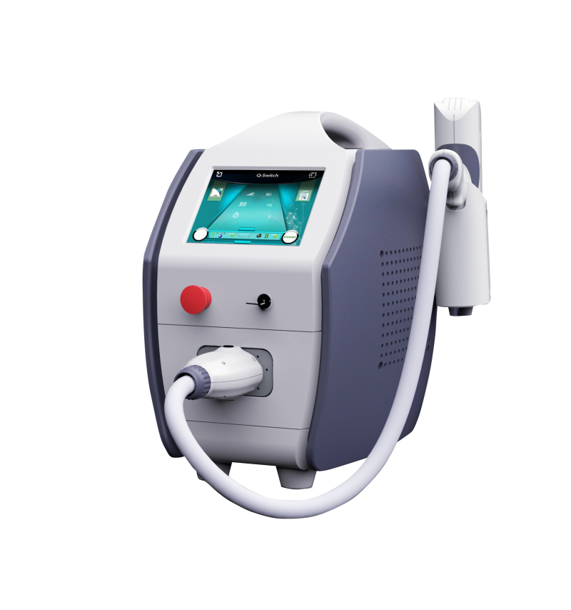
The professional Q Switch Laser available through LorEmA Estetic combines 532 and 1064 nm wavelengths, nanosecond pulses and adjustable spot size. These features support flexible treatment planning but do not replace diagnosis or operator training.
When laser pigment removal is appropriate
Laser treatment has the clearest rationale when the practitioner is treating an assessed benign lesion with a defined target.
Before treatment, review:
-
When the spot appeared and whether it has changed.
-
Possible links to sun exposure, inflammation, pregnancy or medication.
-
Recent tanning.
-
Previous responses to lasers and peels.
-
Photosensitising products or medicines.
-
Features requiring dermatological assessment.
A new, changing, asymmetrical, irregular or multi-coloured lesion should be assessed by a dermatologist. The American Academy of Dermatology’s ABCDE guidance outlines common warning signs.
How to choose a pigmentation device for a clinic
Equipment selection should reflect the clinic’s clients, services and operator competence.
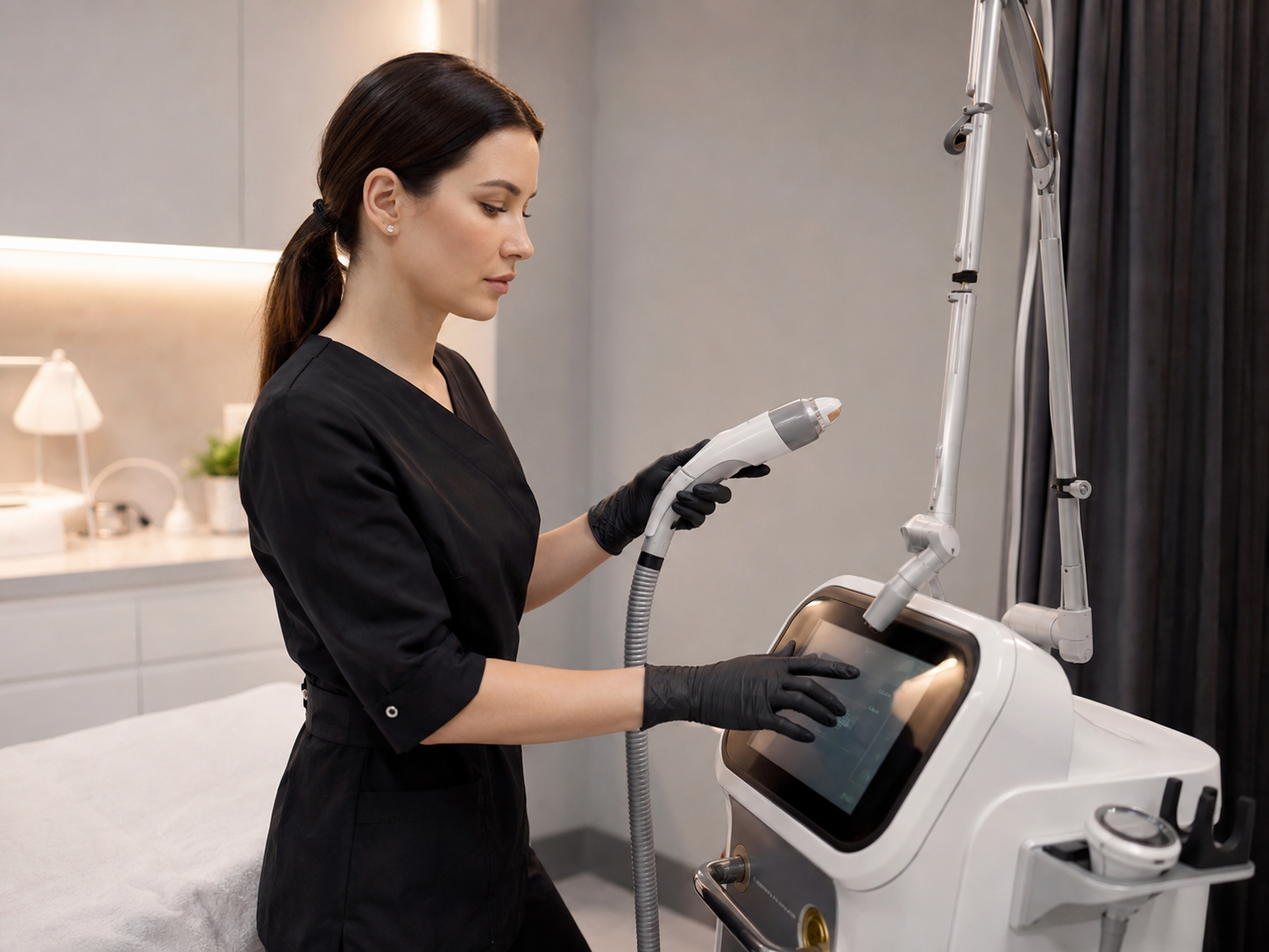
Key criteria include:
-
available wavelengths;
-
adjustable fluence, repetition rate and spot size;
-
pulse stability;
-
cooling and maintenance;
-
training and technical support;
-
potential for service expansion.
A dedicated Q-switched system may suit a practice focused on pigment and tattoo applications. A clinic building a broader laser menu may consider the modular Xlase Plus platform.
Why training matters as much as equipment
A capable device cannot compensate for an incorrect diagnosis. Practitioners need a working understanding of melanogenesis, phototypes, inflammatory responses and light-tissue interaction.
The LorEmA Estetic basic cosmetology course includes skin assessment, dermatological foundations, safety and device-based methods, helping practitioners understand both treatment opportunities and professional limits.
FAQ
Which laser is best for pigmented spots?
There is no universal best device. The correct choice depends on the diagnosis, pigment depth, skin phototype and available parameters.
Is 532 or 1064 nm better?
They serve different purposes. The 532 nm wavelength is used for selected superficial targets, while 1064 nm penetrates more deeply.
Can melasma be removed permanently?
Melasma tends to recur. Procedures may improve its appearance, but ongoing trigger control and photoprotection remain important.
Why can a spot look darker after treatment?
It may be a temporary pigment response or post-inflammatory hyperpigmentation. Risk can increase with tanning, irritation and excessive settings.
Can moles be treated with a cosmetic laser?
Melanocytic and suspicious lesions should be assessed by a dermatologist before any cosmetic treatment.
Which device should a new practice choose?
The choice should reflect operator training, client profile, parameter control, technical support and the services the practice is ready to deliver safely.
Laser selection begins with diagnosis
Pigmented spots differ in origin, depth and behaviour. Professional laser treatment therefore begins with understanding the pigment, not simply choosing the most powerful device.
LorEmA Estetic supports beauty professionals and clinic owners in Moldova with guidance on selecting a professional Q Switch Laser, choosing a wider laser platform and building the knowledge required to introduce pigment treatments responsibly.
